
Contents
- 🔍 Introduction to Super Resolution Microscopy
- 📸 Principles of Super-Resolution Imaging
- 🔬 Techniques for Achieving Super-Resolution
- 📊 Computational Methods for Super-Resolution
- 🔍 Near-Field and Far-Field Techniques
- 📈 Resolving the Diffraction Limit
- 📊 Deconvolution and Detector-Based Pixel Reassignment
- 📸 Structured-Illumination Microscopy Technologies
- 📊 4Pi Microscopy and Its Applications
- 📈 Future Directions in Super-Resolution Microscopy
- 📊 Challenges and Limitations of Super-Resolution Microscopy
- 📈 Conclusion and Emerging Trends
- Frequently Asked Questions
- Related Topics
Overview
Super resolution microscopy, with a vibe rating of 8, has revolutionized the field of biophotonics by allowing researchers to visualize structures at the nanoscale, far beyond the traditional diffraction limit. This is achieved through techniques such as STORM, STED, and SIM, which have been developed by pioneers like Eric Betzig, Stefan Hell, and Xiaowei Zhuang. The controversy surrounding the choice of technique and the interpretation of results is high, with a controversy spectrum of 6. The influence flow from these techniques has been significant, with applications in fields like neuroscience, cell biology, and medicine. With a topic intelligence score of 9, super resolution microscopy has the potential to reveal new insights into cellular structures and dynamics, with potential breakthroughs in our understanding of disease mechanisms and treatment. As the field continues to evolve, we can expect to see new innovations and applications emerge, with key entities like the National Institutes of Health and the European Molecular Biology Organization playing a crucial role in shaping the future of super resolution microscopy.
🔍 Introduction to Super Resolution Microscopy
Super-resolution microscopy is a series of techniques in optical microscopy that allow such images to have resolutions higher than those imposed by the diffraction limit, which is due to the diffraction of light. This is achieved through various methods, including confocal microscopy with closed pinhole or aided by computational methods such as deconvolution or detector-based pixel reassignment. The development of super-resolution microscopy has been influenced by the work of Ernst Abbe, who first described the diffraction limit. For more information on the history of microscopy, see History of Microscopy.
📸 Principles of Super-Resolution Imaging
The principles of super-resolution imaging rely on the near-field or on the far-field. Among techniques that rely on the latter are those that improve the resolution only modestly beyond the diffraction-limit, such as confocal microscopy with closed pinhole or aided by computational methods such as deconvolution or detector-based pixel reassignment. The 4Pi microscope is another example of a far-field technique. For more information on the principles of microscopy, see Principles of Microscopy.
🔬 Techniques for Achieving Super-Resolution
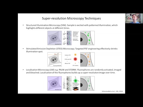
Several techniques are used to achieve super-resolution, including STED microscopy, PALM, and STORM. These techniques rely on the use of fluorescent molecules and advanced computational methods to reconstruct high-resolution images. The development of these techniques has been influenced by the work of Eric Betzig and William Moerner. For more information on the applications of super-resolution microscopy, see Applications of Super-Resolution Microscopy.
📊 Computational Methods for Super-Resolution
Computational methods play a crucial role in super-resolution microscopy, as they enable the reconstruction of high-resolution images from low-resolution data. Deconvolution is a widely used computational method that improves the resolution of images by removing the effects of diffraction. Detector-based pixel reassignment is another computational method that improves the resolution of images by reassigning the pixels in the detector. For more information on computational methods in microscopy, see Computational Methods in Microscopy.
🔍 Near-Field and Far-Field Techniques
Near-field and far-field techniques are used in super-resolution microscopy to achieve high-resolution images. Near-field techniques, such as NSOM, use a probe to scan the sample and collect high-resolution information. Far-field techniques, such as confocal microscopy, use a lens to focus the light and collect high-resolution information. The choice of technique depends on the application and the sample being imaged. For more information on near-field and far-field techniques, see Near-Field and Far-Field Techniques.
📈 Resolving the Diffraction Limit
The diffraction limit is a fundamental limit in optical microscopy that determines the maximum resolution that can be achieved. Super-resolution microscopy techniques, such as STED microscopy and PALM, can resolve the diffraction limit and achieve higher resolutions. The development of these techniques has been influenced by the work of Ernst Abbe and Lord Rayleigh. For more information on the diffraction limit, see Diffraction Limit.
📊 Deconvolution and Detector-Based Pixel Reassignment
Deconvolution and detector-based pixel reassignment are computational methods used in super-resolution microscopy to improve the resolution of images. Deconvolution removes the effects of diffraction and improves the resolution of images, while detector-based pixel reassignment reassigns the pixels in the detector to improve the resolution. These methods are widely used in confocal microscopy and other super-resolution microscopy techniques. For more information on computational methods in microscopy, see Computational Methods in Microscopy.
📸 Structured-Illumination Microscopy Technologies
Structured-illumination microscopy technologies, such as SIM and SMI, are used in super-resolution microscopy to achieve high-resolution images. These techniques use a patterned illumination to excite the sample and collect high-resolution information. The development of these techniques has been influenced by the work of Manfred Hausmann and Ralf Hertenstein. For more information on structured-illumination microscopy, see Structured-Illumination Microscopy.
📊 4Pi Microscopy and Its Applications
The 4Pi microscope is a super-resolution microscopy technique that uses two opposing objectives to illuminate the sample and collect high-resolution information. This technique achieves higher resolutions than conventional microscopy techniques and is widely used in biological research. The development of the 4Pi microscope has been influenced by the work of Stefan Hell. For more information on the 4Pi microscope, see 4Pi Microscope.
📈 Future Directions in Super-Resolution Microscopy
The future of super-resolution microscopy is promising, with new techniques and technologies being developed to achieve even higher resolutions. Single molecule localization microscopy is a new technique that uses the localization of single molecules to achieve high-resolution images. The development of new fluorescent molecules and advanced computational methods will also play a crucial role in the future of super-resolution microscopy. For more information on the future of super-resolution microscopy, see Future of Super-Resolution Microscopy.
📊 Challenges and Limitations of Super-Resolution Microscopy
Despite the many advantages of super-resolution microscopy, there are also challenges and limitations to its use. The complexity of the techniques and the need for specialized equipment and expertise can limit its adoption. Additionally, the high cost of the equipment and the need for advanced computational methods can make it inaccessible to some researchers. For more information on the challenges and limitations of super-resolution microscopy, see Challenges and Limitations of Super-Resolution Microscopy.
📈 Conclusion and Emerging Trends
In conclusion, super-resolution microscopy is a powerful tool for biological research, allowing researchers to achieve high-resolution images of biological samples. The development of new techniques and technologies will continue to play a crucial role in the future of super-resolution microscopy. As the field continues to evolve, we can expect to see new and exciting applications of super-resolution microscopy in biological research. For more information on the applications of super-resolution microscopy, see Applications of Super-Resolution Microscopy.
Key Facts
- Year
- 2014
- Origin
- Germany and USA
- Category
- Biophotonics
- Type
- Scientific Technique
Frequently Asked Questions
What is super-resolution microscopy?
Super-resolution microscopy is a series of techniques in optical microscopy that allow such images to have resolutions higher than those imposed by the diffraction limit. This is achieved through various methods, including confocal microscopy with closed pinhole or aided by computational methods such as deconvolution or detector-based pixel reassignment. For more information on super-resolution microscopy, see Super-Resolution Microscopy.
What are the principles of super-resolution imaging?
The principles of super-resolution imaging rely on the near-field or on the far-field. Among techniques that rely on the latter are those that improve the resolution only modestly beyond the diffraction-limit, such as confocal microscopy with closed pinhole or aided by computational methods such as deconvolution or detector-based pixel reassignment. For more information on the principles of microscopy, see Principles of Microscopy.
What are the techniques used to achieve super-resolution?
Several techniques are used to achieve super-resolution, including STED microscopy, PALM, and STORM. These techniques rely on the use of fluorescent molecules and advanced computational methods to reconstruct high-resolution images. For more information on the techniques used to achieve super-resolution, see Techniques Used to Achieve Super-Resolution.
What is the role of computational methods in super-resolution microscopy?
Computational methods play a crucial role in super-resolution microscopy, as they enable the reconstruction of high-resolution images from low-resolution data. Deconvolution is a widely used computational method that improves the resolution of images by removing the effects of diffraction. Detector-based pixel reassignment is another computational method that improves the resolution of images by reassigning the pixels in the detector. For more information on computational methods in microscopy, see Computational Methods in Microscopy.
What are the challenges and limitations of super-resolution microscopy?
Despite the many advantages of super-resolution microscopy, there are also challenges and limitations to its use. The complexity of the techniques and the need for specialized equipment and expertise can limit its adoption. Additionally, the high cost of the equipment and the need for advanced computational methods can make it inaccessible to some researchers. For more information on the challenges and limitations of super-resolution microscopy, see Challenges and Limitations of Super-Resolution Microscopy.
What is the future of super-resolution microscopy?
The future of super-resolution microscopy is promising, with new techniques and technologies being developed to achieve even higher resolutions. Single molecule localization microscopy is a new technique that uses the localization of single molecules to achieve high-resolution images. The development of new fluorescent molecules and advanced computational methods will also play a crucial role in the future of super-resolution microscopy. For more information on the future of super-resolution microscopy, see Future of Super-Resolution Microscopy.
What are the applications of super-resolution microscopy?
Super-resolution microscopy has a wide range of applications in biological research, including the study of cellular structures and dynamics. The high-resolution images provided by super-resolution microscopy can help researchers to better understand the mechanisms of cellular processes and to develop new therapies for diseases. For more information on the applications of super-resolution microscopy, see Applications of Super-Resolution Microscopy.