
Contents
- 🎵 Origins & History
- ⚙️ How It Works
- 📊 Key Facts & Numbers
- 👥 Key People & Organizations
- 🌍 Cultural Impact & Influence
- ⚡ Current State & Latest Developments
- 🤔 Controversies & Debates
- 🔮 Future Outlook & Predictions
- 💡 Practical Applications
- 📚 Related Topics & Deeper Reading
- Frequently Asked Questions
- References
- Related Topics
Overview
A computed tomography (CT) scan is a medical imaging technique that uses X-rays and computer technology to produce detailed cross-sectional images of the body. Developed in the 1970s, CT scanning has become a versatile tool in medical diagnosis, allowing for the non-invasive examination of internal structures and organs. With its ability to image a wide range of tissues and materials, CT scans have revolutionized the field of medicine, enabling doctors to diagnose and treat various conditions more accurately. According to the National Cancer Institute, over 80 million CT scans are performed annually in the United States alone, highlighting the significance of this technology in modern healthcare. The American College of Radiology also emphasizes the importance of CT scans in patient care, providing guidelines for their safe and effective use. As technology continues to advance, CT scans remain an essential tool in the diagnosis and treatment of diseases, with ongoing research focused on improving image quality, reducing radiation exposure, and exploring new applications in fields like artificial intelligence and personalized medicine.
🎵 Origins & History
The concept of CT scans dates back to the 1960s, when Godfrey Hounsfield, an English engineer, and Allan McLeod Cormack, a South African physicist, independently developed the theoretical foundation for the technology. Their work led to the creation of the first CT scanner in 1971, which was installed at Atkinson Morley Hospital in London. The first patient to undergo a CT scan was a woman with a suspected brain tumor, and the resulting images provided unprecedented detail and clarity, marking the beginning of a new era in medical imaging. Since then, CT scanning has undergone significant advancements, with improvements in image resolution, scan time, and radiation dose. The introduction of helical CT in the 1990s, for example, enabled the scanning of larger areas of the body in a single rotation, increasing the speed and efficiency of the process.
⚙️ How It Works
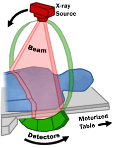
CT scanners use a rotating X-ray tube and a row of detectors placed in a gantry to measure X-ray attenuations by different tissues inside the body. The multiple X-ray measurements taken from different angles are then processed on a computer using tomographic reconstruction algorithms to produce tomographic (cross-sectional) images (virtual 'slices') of a body. This process allows for the creation of detailed images of internal structures, including bones, organs, and soft tissues. The Food and Drug Administration (FDA) regulates the safety and efficacy of CT scanners, ensuring that they meet strict standards for image quality and radiation exposure. Additionally, the National Institute of Biomedical Imaging and Bioengineering (NIBIB) supports research and development of new CT technologies, including the use of artificial intelligence and machine learning to improve image analysis and diagnosis.
📊 Key Facts & Numbers
CT scans have numerous applications in medical diagnosis, including the detection of tumors, vascular diseases, and internal injuries. According to the Centers for Disease Control and Prevention (CDC), CT scans are used in over 50% of all emergency department visits, highlighting their importance in acute care. The American College of Radiology (ACR) provides guidelines for the appropriate use of CT scans, emphasizing the need for careful consideration of radiation exposure and image quality. Furthermore, the National Cancer Institute (NCI) supports research on the use of CT scans in cancer diagnosis and treatment, including the development of new imaging agents and techniques to improve detection and monitoring of tumors.
👥 Key People & Organizations
Key people involved in the development and advancement of CT scanning include Godfrey Hounsfield and Allan McLeod Cormack, who were awarded the Nobel Prize in Physiology or Medicine in 1979 for their contributions to the field. Other notable organizations involved in CT scanning include the American College of Radiology (ACR), the National Institute of Biomedical Imaging and Bioengineering (NIBIB), and the Food and Drug Administration (FDA). The International Society for Computed Tomography (ISCT) also plays a crucial role in promoting research, education, and innovation in the field of CT scanning.
🌍 Cultural Impact & Influence
CT scans have had a significant impact on modern medicine, enabling doctors to diagnose and treat a wide range of conditions more accurately and effectively. The technology has also led to the development of new medical specialties, such as interventional radiology, which involves the use of imaging guidance to perform minimally invasive procedures. The World Health Organization (WHO) recognizes the importance of CT scans in global healthcare, providing guidelines for their use in low- and middle-income countries. Additionally, the National Institutes of Health (NIH) supports research on the use of CT scans in various diseases, including cancer, cardiovascular disease, and neurological disorders.
⚡ Current State & Latest Developments
Current research and developments in CT scanning focus on improving image quality, reducing radiation exposure, and exploring new applications in fields like artificial intelligence and personalized medicine. The introduction of spectral CT and photon-counting CT has enabled the creation of more detailed and quantitative images, while advances in machine learning and deep learning are being explored for their potential to improve image analysis and diagnosis. The American College of Radiology (ACR) and the National Institute of Biomedical Imaging and Bioengineering (NIBIB) are at the forefront of these developments, supporting research and innovation in the field of CT scanning.
🤔 Controversies & Debates
Despite the many benefits of CT scans, there are also controversies and debates surrounding their use, particularly with regards to radiation exposure and the potential risks associated with it. The National Cancer Institute (NCI) and the Food and Drug Administration (FDA) have issued guidelines and warnings about the risks of radiation exposure from CT scans, emphasizing the need for careful consideration and justification of their use. The American College of Radiology (ACR) also provides guidelines for the safe and effective use of CT scans, including recommendations for radiation dose reduction and image quality improvement.
🔮 Future Outlook & Predictions
Looking to the future, CT scans are likely to continue playing a vital role in medical diagnosis and treatment, with ongoing research and development focused on improving image quality, reducing radiation exposure, and exploring new applications in fields like artificial intelligence and personalized medicine. The introduction of new technologies, such as quantum CT and nanoparticle-enhanced CT, is expected to further enhance the capabilities of CT scanning, enabling doctors to diagnose and treat diseases more accurately and effectively. The National Institutes of Health (NIH) and the National Institute of Biomedical Imaging and Bioengineering (NIBIB) will continue to play a crucial role in supporting research and innovation in the field of CT scanning.
💡 Practical Applications
CT scans have numerous practical applications in medical diagnosis and treatment, including the detection of tumors, vascular diseases, and internal injuries. They are also used in interventional radiology to guide minimally invasive procedures, such as biopsies and tumor treatments. The American College of Radiology (ACR) provides guidelines for the appropriate use of CT scans, emphasizing the need for careful consideration of radiation exposure and image quality. Additionally, the National Cancer Institute (NCI) supports research on the use of CT scans in cancer diagnosis and treatment, including the development of new imaging agents and techniques to improve detection and monitoring of tumors.
Key Facts
- Year
- 1971
- Origin
- London, UK
- Category
- public-health
- Type
- technology
Frequently Asked Questions
What is a CT scan?
A CT scan is a medical imaging technique that uses X-rays and computer technology to produce detailed cross-sectional images of the body. It is commonly used to diagnose and treat a wide range of medical conditions, including tumors, vascular diseases, and internal injuries. According to the National Cancer Institute, CT scans are used in over 50% of all emergency department visits. The American College of Radiology (ACR) provides guidelines for the safe and effective use of CT scans, emphasizing the need for careful consideration of radiation exposure and image quality.
How does a CT scan work?
A CT scan works by using a rotating X-ray tube and a row of detectors placed in a gantry to measure X-ray attenuations by different tissues inside the body. The multiple X-ray measurements taken from different angles are then processed on a computer using tomographic reconstruction algorithms to produce tomographic (cross-sectional) images (virtual 'slices') of a body. The Food and Drug Administration (FDA) regulates the safety and efficacy of CT scanners, ensuring that they meet strict standards for image quality and radiation exposure.
What are the benefits of CT scans?
CT scans have numerous benefits, including the ability to diagnose and treat a wide range of medical conditions, such as tumors, vascular diseases, and internal injuries. They are also non-invasive and relatively quick, making them a convenient option for patients. The National Institutes of Health (NIH) supports research on the use of CT scans in various diseases, including cancer, cardiovascular disease, and neurological disorders. Additionally, the American College of Radiology (ACR) provides guidelines for the appropriate use of CT scans, emphasizing the need for careful consideration of radiation exposure and image quality.
What are the risks associated with CT scans?
CT scans involve exposure to X-rays, which can increase the risk of cancer and other health problems. The National Cancer Institute (NCI) and the Food and Drug Administration (FDA) have issued guidelines and warnings about the risks of radiation exposure from CT scans, emphasizing the need for careful consideration and justification of their use. The American College of Radiology (ACR) also provides guidelines for the safe and effective use of CT scans, including recommendations for radiation dose reduction and image quality improvement.
How are CT scans used in medical diagnosis and treatment?
CT scans are used in medical diagnosis and treatment to detect and diagnose a wide range of medical conditions, including tumors, vascular diseases, and internal injuries. They are also used to guide minimally invasive procedures, such as biopsies and tumor treatments. The American College of Radiology (ACR) provides guidelines for the appropriate use of CT scans, emphasizing the need for careful consideration of radiation exposure and image quality. Additionally, the National Cancer Institute (NCI) supports research on the use of CT scans in cancer diagnosis and treatment, including the development of new imaging agents and techniques to improve detection and monitoring of tumors.
What is the future of CT scans?
The future of CT scans is likely to involve ongoing research and development focused on improving image quality, reducing radiation exposure, and exploring new applications in fields like artificial intelligence and personalized medicine. The introduction of new technologies, such as quantum CT and nanoparticle-enhanced CT, is expected to further enhance the capabilities of CT scanning, enabling doctors to diagnose and treat diseases more accurately and effectively. The National Institutes of Health (NIH) and the National Institute of Biomedical Imaging and Bioengineering (NIBIB) will continue to play a crucial role in supporting research and innovation in the field of CT scanning.
How do CT scans compare to other medical imaging technologies?
CT scans have their own unique advantages and limitations compared to other medical imaging technologies, such as magnetic resonance imaging (MRI) and positron emission tomography (PET). CT scans are often used in combination with these technologies to provide a more comprehensive understanding of the body. The International Society for Magnetic Resonance in Medicine (ISMRM) and the Society of Nuclear Medicine and Molecular Imaging (SNMMI) are prominent organizations in the field of medical imaging, promoting research, education, and innovation in these areas.
What are the potential applications of CT scans in personalized medicine?
CT scans have the potential to play a significant role in personalized medicine, enabling doctors to tailor treatment plans to individual patients based on their unique characteristics and needs. The National Institutes of Health (NIH) supports research on the use of CT scans in personalized medicine, including the development of new imaging agents and techniques to improve detection and monitoring of diseases. Additionally, the American College of Radiology (ACR) provides guidelines for the appropriate use of CT scans in personalized medicine, emphasizing the need for careful consideration of radiation exposure and image quality.