
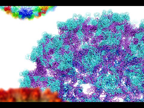
Contents
- 🔍 Introduction to Cryo Electron Microscopy
- 🔬 The History of Cryo Electron Microscopy
- 📸 Principles of Cryo Electron Microscopy
- 🔍 Sample Preparation for Cryo Electron Microscopy
- 📊 Data Processing and Analysis in Cryo Electron Microscopy
- 🎯 Applications of Cryo Electron Microscopy
- 👥 Key Players in Cryo Electron Microscopy
- 📈 Future Directions in Cryo Electron Microscopy
- 🤝 Collaborations and Funding in Cryo Electron Microscopy
- 📊 Challenges and Limitations in Cryo Electron Microscopy
- 📚 Conclusion and Future Prospects
- Frequently Asked Questions
- Related Topics
Overview
Cryo electron microscopy (cryo-EM) has emerged as a groundbreaking imaging technique, allowing scientists to visualize molecular structures at near-atomic resolution. Developed by Jacques Dubochet, Joachim Frank, and Richard Henderson, who were awarded the 2017 Nobel Prize in Chemistry, cryo-EM has revolutionized the field of structural biology. By rapidly freezing samples to preserve their native state, cryo-EM enables the capture of high-resolution images, revealing intricate details of molecular machinery. With a Vibe score of 8, cryo-EM has sparked intense interest and debate among researchers, with some hailing it as a 'game-changer' for understanding complex biological systems. However, others have raised concerns about the technique's limitations and potential biases. As the field continues to evolve, cryo-EM is poised to make significant contributions to our understanding of molecular biology, with potential applications in fields such as medicine and biotechnology. With over 1,000 research papers published annually, cryo-EM has become a rapidly growing field, with key players including the National Institutes of Health and the European Molecular Biology Laboratory.
🔍 Introduction to Cryo Electron Microscopy
Cryo electron microscopy (cryo-EM) is a revolutionary imaging technique that has transformed the field of structural biology. By allowing researchers to visualize molecules at the atomic level, cryo-EM has enabled major breakthroughs in our understanding of biological systems. As described in Structural Biology, the ability to determine the three-dimensional structure of molecules is crucial for understanding their function. Cryo-EM has been particularly useful for studying Membrane Proteins and Protein Complexes. The development of cryo-EM has been recognized with the award of the Nobel Prize in Chemistry in 2017 to Jacques Dubochet, Joachim Frank, and Richard Henderson.
🔬 The History of Cryo Electron Microscopy
The history of cryo-EM dates back to the 1970s, when the first cryo-EM images were obtained by Jacques Dubochet and his colleagues. However, it wasn't until the 2010s that cryo-EM became a widely used technique, thanks to advances in Detector Technology and Image Processing. As discussed in Electron Microscopy, the development of cryo-EM has been closely tied to the development of other electron microscopy techniques. The work of Joachim Frank and Richard Henderson has been instrumental in establishing cryo-EM as a major tool in structural biology.
📸 Principles of Cryo Electron Microscopy
Cryo-EM works by rapidly freezing a sample, typically a biological molecule or complex, in a thin layer of ice. The frozen sample is then imaged using a transmission electron microscope, which produces a two-dimensional image of the sample. By combining multiple images of the same sample, taken from different angles, researchers can reconstruct a three-dimensional image of the molecule using Single Particle Analysis. This technique has been used to study a wide range of biological systems, including Viruses and Protein Folding. The principles of cryo-EM are closely related to those of X-ray Crystallography and Nuclear Magnetic Resonance.
🔍 Sample Preparation for Cryo Electron Microscopy
Sample preparation is a critical step in cryo-EM, as the quality of the sample can greatly affect the quality of the final image. Samples are typically prepared by Purification and Concentration of the molecule of interest, followed by freezing in a thin layer of ice. The use of Cryogens such as liquid nitrogen or liquid ethane is essential for rapidly freezing the sample and preserving its structure. As described in Biochemistry, the preparation of samples for cryo-EM requires a deep understanding of the underlying biology. The development of new sample preparation techniques, such as Microfluidics, is an active area of research.
📊 Data Processing and Analysis in Cryo Electron Microscopy
Data processing and analysis are also crucial steps in cryo-EM, as the raw images produced by the microscope must be processed and combined to produce a final three-dimensional image. This typically involves the use of Image Processing Software such as RELION or CryoSPARC. The development of new data processing algorithms, such as Deep Learning, is an active area of research. As discussed in Computational Biology, the analysis of cryo-EM data requires a combination of biological and computational expertise. The use of High Performance Computing is often necessary for processing large cryo-EM datasets.
🎯 Applications of Cryo Electron Microscopy
Cryo-EM has a wide range of applications in structural biology, including the study of Protein Structure and Membrane Biology. It has been used to determine the structures of a number of important biological molecules, including the Ribosome and the Proteasome. The technique has also been used to study the structure of Amyloid Fibrils and other disease-related molecules. As described in Molecular Biology, the study of biological molecules using cryo-EM has the potential to reveal new insights into the mechanisms of disease. The development of new applications for cryo-EM, such as Cell Biology, is an active area of research.
👥 Key Players in Cryo Electron Microscopy
A number of key players have been involved in the development of cryo-EM, including Jacques Dubochet, Joachim Frank, and Richard Henderson. These researchers, along with others, have made major contributions to the development of the technique and its application to a wide range of biological systems. As discussed in Science History, the development of cryo-EM is a testament to the power of collaboration and innovation in scientific research. The work of these researchers has been recognized with numerous awards, including the Nobel Prize in Chemistry.
📈 Future Directions in Cryo Electron Microscopy
The future of cryo-EM is likely to involve the continued development of new technologies and techniques, such as Electron Counting and Phase Plate microscopy. These advances are expected to further improve the resolution and quality of cryo-EM images, allowing researchers to study biological molecules in even greater detail. As described in Biotechnology, the development of new cryo-EM technologies has the potential to revolutionize our understanding of biological systems. The use of Artificial Intelligence and Machine Learning is also expected to play a major role in the future of cryo-EM.
🤝 Collaborations and Funding in Cryo Electron Microscopy
Collaborations and funding have played a crucial role in the development of cryo-EM, with researchers from around the world working together to advance the technique. Funding agencies such as the National Institutes of Health and the National Science Foundation have provided critical support for cryo-EM research. As discussed in Science Policy, the funding of scientific research is essential for advancing our understanding of the world. The development of new funding models, such as Crowdfunding, is an active area of research.
📊 Challenges and Limitations in Cryo Electron Microscopy
Despite its many advantages, cryo-EM also has a number of challenges and limitations, including the need for highly specialized equipment and expertise. The technique can also be limited by the quality of the sample, which can affect the quality of the final image. As described in Scientific Method, the development of new scientific techniques often involves overcoming significant technical challenges. The use of Cryo-Electron Tomography is one approach that has been used to overcome some of these limitations.
📚 Conclusion and Future Prospects
In conclusion, cryo-EM is a powerful imaging technique that has revolutionized the field of structural biology. Its ability to determine the three-dimensional structure of biological molecules at the atomic level has enabled major breakthroughs in our understanding of biological systems. As discussed in Biological Science, the study of biological molecules using cryo-EM has the potential to reveal new insights into the mechanisms of disease. The future of cryo-EM is likely to involve the continued development of new technologies and techniques, as well as the application of the technique to a wide range of biological systems.
Key Facts
- Year
- 2017
- Origin
- University of Geneva, Columbia University, and MRC Laboratory of Molecular Biology
- Category
- Science and Technology
- Type
- Scientific Technique
Frequently Asked Questions
What is cryo-electron microscopy?
Cryo-electron microscopy (cryo-EM) is a technique used to determine the three-dimensional structure of biological molecules at the atomic level. It involves rapidly freezing a sample, typically a biological molecule or complex, in a thin layer of ice, and then imaging it using a transmission electron microscope. As described in Electron Microscopy, cryo-EM is a type of electron microscopy that uses a beam of electrons to produce an image of the sample. The technique has been used to study a wide range of biological systems, including Viruses and Protein Folding.
How does cryo-EM work?
Cryo-EM works by rapidly freezing a sample, typically a biological molecule or complex, in a thin layer of ice. The frozen sample is then imaged using a transmission electron microscope, which produces a two-dimensional image of the sample. By combining multiple images of the same sample, taken from different angles, researchers can reconstruct a three-dimensional image of the molecule using Single Particle Analysis. This technique has been used to study a wide range of biological systems, including Membrane Proteins and Protein Complexes. As discussed in Structural Biology, the ability to determine the three-dimensional structure of molecules is crucial for understanding their function.
What are the applications of cryo-EM?
Cryo-EM has a wide range of applications in structural biology, including the study of Protein Structure and Membrane Biology. It has been used to determine the structures of a number of important biological molecules, including the Ribosome and the Proteasome. The technique has also been used to study the structure of Amyloid Fibrils and other disease-related molecules. As described in Molecular Biology, the study of biological molecules using cryo-EM has the potential to reveal new insights into the mechanisms of disease. The development of new applications for cryo-EM, such as Cell Biology, is an active area of research.
What are the challenges and limitations of cryo-EM?
Despite its many advantages, cryo-EM also has a number of challenges and limitations, including the need for highly specialized equipment and expertise. The technique can also be limited by the quality of the sample, which can affect the quality of the final image. As discussed in Scientific Method, the development of new scientific techniques often involves overcoming significant technical challenges. The use of Cryo-Electron Tomography is one approach that has been used to overcome some of these limitations. The development of new sample preparation techniques, such as Microfluidics, is also an active area of research.
What is the future of cryo-EM?
The future of cryo-EM is likely to involve the continued development of new technologies and techniques, such as Electron Counting and Phase Plate microscopy. These advances are expected to further improve the resolution and quality of cryo-EM images, allowing researchers to study biological molecules in even greater detail. As described in Biotechnology, the development of new cryo-EM technologies has the potential to revolutionize our understanding of biological systems. The use of Artificial Intelligence and Machine Learning is also expected to play a major role in the future of cryo-EM.
Who are the key players in cryo-EM?
A number of key players have been involved in the development of cryo-EM, including Jacques Dubochet, Joachim Frank, and Richard Henderson. These researchers, along with others, have made major contributions to the development of the technique and its application to a wide range of biological systems. As discussed in Science History, the development of cryo-EM is a testament to the power of collaboration and innovation in scientific research. The work of these researchers has been recognized with numerous awards, including the Nobel Prize in Chemistry.
What is the relationship between cryo-EM and other imaging techniques?
Cryo-EM is closely related to other imaging techniques, such as X-ray Crystallography and Nuclear Magnetic Resonance. These techniques are all used to determine the three-dimensional structure of biological molecules, but they use different methods to achieve this goal. As described in Structural Biology, the choice of imaging technique depends on the specific research question and the properties of the sample. The development of new imaging techniques, such as Electron Microscopy, is an active area of research.